Table of contents
- Vitamin D deficiency and vitamin D receptor FokI polymorphism as risk factors for COVID-19
- Vitamin D Life -
36 studies in both categories Virus and VDR - Vitamin D Life – COVID-19 treated by Vitamin D - studies, reports, videos
- Vitamin D Life - Vitamin D Receptor activation can be increased in many ways
- Diseases associated with a poor Vitamin D Receptor
Vitamin D deficiency and vitamin D receptor FokI polymorphism as risk factors for COVID-19
Pediatr Res. 2022 Sep 9. doi: 10.1038/s41390-022-02275-6
Nancy M S Zeidan 1 , Hanan M Abd El Lateef 2 , Dalia M Selim 2 , Suzan A Razek 2 , Ghada A B Abd-Elrehim 3 , Mohamed Nashat 4 , Noha ElGyar 5 , Nevin M Waked 6 , Attia A Soliman 7 , Ahmed A Elhewala 7 , Mohamed M M Shehab 7 , Ahmed A A Ibraheem 7 , Hassan Shehata 7 , Yousif M Yousif 7 , Nagwa E Akeel 7 , Mustafa I A Hashem 7 , Amani A Ahmed 7 , Ahmed A Emam 8 , Mohamed M Abdelmohsen 9 , Mohamed F Ahmed 9 , Ahmed S E Saleh 10 , Heba H Eltrawy 11 , Gehan H Shahin 12 , Rehab M Nabil 13 , Thoraya A Hosny 13 , Mohamed R Abdelhamed 14 , Mona R Afify 15 , Mohanned T Alharbi 15 , Mohammed K Nagshabandi 15 , Muyassar K Tarabulsi 15 , Sherif F Osman 16 , Amal S M Abd-Elrazek 17 , Manal M Rashad 18 , Sonya A A El-Gaaly 19 , Said A B Gad 20 , Mohamed Y Mohamed 21 , Khalil Abdelkhalek 1 , Aly A Yousef 22Background: Given the sparse data on vitamin D status in pediatric COVID-19, we investigated whether vitamin D deficiency could be a risk factor for susceptibility to COVID-19 in Egyptian children and adolescents. We also investigated whether vitamin D receptor (VDR) FokI polymorphism could be a genetic marker for COVID-19 susceptibility.
Methods: One hundred and eighty patients diagnosed to have COVID-19 and 200 matched control children and adolescents were recruited. Patients were laboratory confirmed as SARS-CoV-2 positive by real-time RT-PCR. All participants were genotyped for VDR Fok1 polymorphism by RT-PCR. Vitamin D status was defined as sufficient for serum 25(OH) D at least 30 ng/mL, insufficient at 21-29 ng/mL, deficient at <20 ng/mL.
Results: Ninety-four patients (52%) had low vitamin D levels with 74 (41%) being deficient and 20 (11%) had vitamin D insufficiency. Vitamin D deficiency was associated with 2.6-fold increased risk for COVID-19 (OR = 2.6; [95% CI 1.96-4.9]; P = 0.002. The FokI FF genotype was significantly more represented in patients compared to control group (OR = 4.05; [95% CI: 1.95-8.55]; P < 0.001).
Conclusions: Vitamin D deficiency and VDR Fok I polymorphism may constitute independent risk factors for susceptibility to COVID-19 in Egyptian children and adolescents.
Impact: Vitamin D deficiency could be a modifiable risk factor for COVID-19 in children and adolescents because of its immune-modulatory action. To our knowledge, ours is the first such study to investigate the VDR Fok I polymorphism in Caucasian children and adolescents with COVID-19. Vitamin D deficiency and the VDR Fok I polymorphism may constitute independent risk factors for susceptibility to COVID-19 in Egyptian children and adolescents. Clinical trials should be urgently conducted to test for causality and to evaluate the efficacy of vitamin D supplementation for prophylaxis and treatment of COVID-19 taking into account the VDR polymorphisms.
Case definition
One hundred and eighty patients aged <19 years who were diagnosed to have COVID-19 at the study hospitals were recruited. All patients were laboratory confirmed as SARS-CoV-2 positive by real-time reverse transcriptase polymerase chain reaction (RT-PCR) assay of nasopharyngeal swab specimens.
Patients' COVID-19 illness severity was categorized into moderate, severe, and critical subgroups according to the recently published classification by Chen et al.20
No asymptomatic or mild cases were seen among our cohort.- (I) Moderate: included (134) cases presented with pneumonia (lower respiratory symptoms plus fever >38 °C and age-specific tachypnea);
- (II) Severe COVID-19: included (31) cases who rapidly develop dyspnea, hypoxia (arterial oxygen saturation <93%), dehydration with feeding difficulty, elevated liver enzymes, disturbed consciousness, coagulation dysfunction;
- (III) Critically ill cases: included (15) patients who required intensive care unit (ICU) monitoring for acute respiratory failure (ARF), mechanical ventilation (partial arterial oxygen pressure/fraction of inspired oxygen PaO2/FiO2 ratio <300 despite oxygen therapy), septic shock, or organ failure.
Patients were admitted within 72 h from onset of fever and cough. Pulmonary high-resolution computed tomographic images were routinely performed for all patients and evaluated by two experienced radiologists (S.F.O. and A.S.M.) who were blinded to the patients' clinical data.
Control group
Two hundred healthy children and adolescents of matched age, sex, and season at enrollment who underwent pre-operative assessment for elective surgery at the study hospitals were enrolled as a control group (all tested negative for SARS-Cov2 by RT-PCR and had negative anti-N antibodies test for SARS-Cov2).
All patients and control subjects belong to the same ethnic group: African Caucasian.Exclusion criteria
Patients with obesity, malnutrition, immunodeficiency, congenital heart disease, malignancy, metabolic diseases, autoimmune disorders, or any chronic debilitating disease were excluded. Those who received vitamin D, calcium, multi-vitamin, or mineral supplementation during the previous 6 months were also excluded.
Download the PDF from Vitamin D LifeREFERENCES
- World Health Organization. WHO coronavirus (COVID-19) dashboard. https:// covid19.who.int (2022).
- Huang, C. et al. Clinical features of patients infected with 2019 novel coronavirus in Wuhan, China. Lancet 395, 497-506 (2020).
- Hoffmann, M. et al. SARS-CoV-2 cell entry depends on ACE2 and TMPRSS2 and is blocked by a clinically proven protease inhibitor. Cell 181, 271.e8-280.e8 (2020).
- Guan, W. J. et al. Clinical characteristics of coronavirus disease 2019 in China. N. Engl. J. Med. 382, 1708-1720 (2020).
- Wang, D. et al. Clinical characteristics of 138 hospitalized patients with 2019 novel coronavirus-infected pneumonia in Wuhan, China. JAMA 323, 1061-1069 (2020).
- Pike, J. W. & Meyer, M. B. The vitamin D receptor: new paradigms for the regulation of gene expression by 1,25-dihydroxyvitamin D(3). Endocrinol. Metab. Clin. North Am. 39, 255-269 (2010).
- Martineau, A. R. et al. Vitamin D supplementation to prevent acute respiratory tract infections: systematic review and meta-analysis of individual participant data. BMJ 356, i6583 (2017).
- Dancer, R. C. et al. Vitamin D deficiency contributes directly to the acute respiratory distress syndrome (ARDS). Thorax 70, 617-624 (2015).
- Underwood, M. A. & Bevins, C. L. Defensin-barbed innate immunity: clinical associations in the pediatric population. Pediatrics 125, 1237-1247 (2010).
- Long, Q. X. et al. Clinical and immunological assessment of asymptomatic SARS- CoV-2 infections. Nat. Med. 26, 845-848 (2020).
- Grant, W. B. et al. Evidence that vitamin D supplementation could reduce risk of influenza and COVID-19 infections and deaths. Nutrients 12, 988 (2020).
- Li, Y., Li, Q., Zhang, N. & Liu, Z. Sunlight and vitamin D in the prevention of coronavirus disease (COVID-19) infection and mortality in the United States. Preprint at https://doi.org/10.21203/rs3.rs-32499/v1 (2020).
- Daneshkhah, A. et al. Evidence for possible association of vitamin D status with cytokine storm and unregulated inflammation in COVID-19 patients. Aging Clin. Exp. Res. 32, 2141 -2158 (2020).
- Wang, Q., Xi, B., Reilly, K. H., Liu, M. & Fu, M. Quantitative assessment of the associations between four polymorphisms (FokI, ApaI, BsmI, TaqI) of vitamin D receptor gene and risk of diabetes mellitus. Mol. Biol. Rep. 39, 9405-9414 (2012).
- Neill, O. et al. Vitamin D receptor gene expression and function in a South African population: ethnicity, vitamin D and FokI. PLoS ONE 8, e67663 (2013).
- van Etten, E. et al. The vitamin D receptor gene FokI polymorphism: functional impact on the immune system. Eur. J. Immunol. 37, 395-405 (2007).
- Yilmaz, K. & $en, V. Is vitamin D deficiency a risk factor for COVID-19 in children? Pediatr. Pulmonol. 55, 3595-3601 (2020).
- Alpcan, A., Tursun, S. & Kandur, Y. Vitamin D levels in children with COVID-19: a report from Turkey. Epidemiol. Infect. 149, E180 (2021).
- Akoglu, H. A. et al. Evaluation of childhood COVID-19 cases: a retrospective analysis. J. Pediatr. Infect. Dis. 16, 091-098 (2021).
- Chen, Z. M. et al. Diagnosis and treatment recommendations for pediatric respiratory infection caused by the 2019 novel coronavirus. World J. Pediatr. 16, 240-246 (2020).
- Holick, M. F. The vitamin D deficiency pandemic: approaches for diagnosis, treatment and prevention. Rev. Endocr. Metab. Disord. 18, 153-165 (2017).
- Freire-Paspuel, B. et al. Analytical and clinical comparison of Viasure (CerTest Biotec) and 2019-nCoV CDC (IDT) RT-qPCR kits for SARS-CoV2 diagnosis. Virology 553, 154-156 (2021).
- Fedirko, V. et al. Pre-diagnostic 25-hydroxyvitamin D, VDR and CASR polymorphisms and survival in patients with colorectal cancer in Western European Populations. Cancer Epidemiol. Biomark. Prev. 21, 582-593 (2012).
- Li, W. et al. Association between serum 25-hydroxyvitamin D concentration and pulmonary infection in children. Medicine 97, e9060 (2018).
- McNally, J. D. et al. Vitamin D deficiency in young children with severe acute lower respiratory infection. Pediatr. Pulmonol. 44, 981-988 (2009).El
- Basha, N., Mohsen, M., Kamal, M. & Mehaney, D. Association of vitamin D deficiency with severe pneumonia in hospitalized children under 5 years. Comp. Clin. Pathol. 23, 1247-1252 (2014).
- Chiodini, I. et al. Vitamin D status and SARS-CoV-2 infection and COVID-19 clinical outcomes. Front Public Health 9, 736665 (2021).
- Merzon, E. et al. Low plasma 25(OH) vitamin D level is associated with increased risk of COVID-19 infection: an Israeli population-based study. FEBS J. 287, 3693-3702 (2020).
- Pinzon, R. T. & Pradana, A. W. Vitamin D deficiency among patients with COVID- 19: case series and recent literature review. Trop. Med. Health 48, 102 (2020).
- Beyazgul, G. et al. How vitamin D levels of children changed during COVID-19 pandemic: a comparison of pre-pandemic and pandemic periods. J. Clin. Res. Pediatr. Endocrinol. 14, 188-195 (2022).
- Darren, A. et al. Vitamin D status of children with paediatric inflammatory multisystem syndrome temporally associated with severe acute respiratory syndrome coronavirus 2 (PIMS-TS). Br. J. Nutr. 127, 896-903 (2022).
- Carpagnano, G. E. et al. Vitamin D deficiency as a predictor of poor prognosis in patients with acute respiratory failure due to COVID-19. J. Endocrinol. Investig. 44, 765-771 (2020).
- de Souza, T. H., Nadal, J. A., Nogueira, R. J. N., Pereira, R. M. & Brandao, M. B. Clinical manifestations of children with COVID-19: a systematic review. Pediatr. Pulmonol. 55, 1892-1899 (2020).
- Kong, J. et al. VDR attenuates acute lung injury by blocking Ang-2-Tie-2 pathway and renin-angiotensin system. Mol. Endocrinol. 27, 2116-2125 (2013).
- Greiller, C. L. & Martineau, A. R. Modulation of the immune response to respiratory viruses by vitamin D. Nutrients 7, 4240-4270 (2015).
- Yuan, W. et al. 1, 25-dihydroxyvitamin D3 suppresses renin gene transcription by blocking the activity of the cyclic AMP response element in the renin gene promoter. J. Biol. Chem. 282, 29821 -29830 (2007).
- Overbergh, L. et al. 1alpha,25-dihydroxyvitamin D3 induces an autoantigen- specific T-helper 1/T-helper 2 immune shift in NOD mice immunized with GAD65 (p524-543). Diabetes 49, 1301-1307 (2000).
- Shafiek, H. K. et al. Cytokine profile in Egyptian children and adolescents with COVID-19 pneumonia: a multicenter study. Pediatr. Pulmonol. 56, 3924-3933 (2021).
- Kresfelder, T. L., Janssen, R., Bont, L., Pretorius, M. & Venter, M. Confirmation of an association between single nucleotide polymorphisms in the VDR gene with respiratory syncytial virus related disease in South African children. J. Med. Virol.1834-1840 (2011).
- Han, W. G. et al. Association of vitamin D receptor polymorphism with susceptibility to symptomatic pertussis. PLoS ONE 11, e0149576 (2016).
- Li, W. et al. Polymorphism rs2239185 in vitamin D receptor gene is associated with severe community-acquired pneumonia of children in Chinese Han population: a case-control study. Eur. J. Pediatr. 174, 621-629 (2015).
- Abouzeid, H. et al. Association of vitamin D receptor gene FokI polymorphism and susceptibility to CAP in Egyptian children: a multicenter study. Pediatr. Res. 639-644 (2018).
- Apaydin, T. et al. Effects of vitamin D receptor gene polymorphisms on the prognosis of COVID-19. Clin. Endocrinol. 96, 819-830 (2022).
- Abdollahzadeh, R. et al. Association of Vitamin D receptor gene polymorphisms and clinical/severe outcomes ofCOVID-19 patients. Infect. Genet. Evol. 96,105098 (2021).
- Jurutka, P. W. et al. The polymorphic N terminus in human vitamin D receptor isoforms influences transcriptional activity by modulating interaction with transcription factor IIB. Mol.
Vitamin D Life -
36 studies in both categories Virus and VDR This list is automatically updated
- COVID maximum downregulation of Vitamin D receptor and CYP27B1 resulted in death - Feb 2024
- COVID in hospital stopped by Vitamin D Receptor activators (curcumin, quercetin) – RCT June 2023
- Children with COVID 4X more likely to have poor Vitamin D Receptors (Note: COVID deactivates VDR) – April 2023
- Diabetes 3X more likely if had COVID ICU (VDR was deactivated) - April 2023
- COVID variants protect themselves by deactivating different VDR variants– March 2023
- Dengue Fever decimated by Vitamin D - many studies
- COVID kids were more likely to have a poor VDR (4.3 X), than low Vitamin D (2.6 X) – Sept 2022
- Cancers are associated with low vitamin D, poor vaccination response and perhaps poor VDR – July 2022
- COVID 3X more likely if a poor Receptor (cells get less Vitamin D from the blood) – July 2022
- Long-COVID is now the biggest COVID concern - many studies
- COVID death 12X more likely if poor Vitamin D Receptor (less D gets to cells) - many studies
- COVID severity, ICU, and mortality all associated with poor vitamin D receptor (but not D, everyone had low D) -Dec 2021
- Different Vitamin D Receptor problems cause different COVID problems - Dec 2021
- COVID-19 severity associated with 3 vitamin D genes – Oct 2021
- Poor Vitamin D receptor blocked Vitamin D from fighting avian influenza viruses (in mice) – July 2021
- Epstein-Barr is yet another virus that deactivates the Vitamin D receptor (COVID later suspected as well)– 2010
- COVID-19 symptoms and comorbidities associated with the type of Vitamin D Receptor – Oct 2021
- Enveloped virus infection (RSV, coronavirus, HIV, etc.) 1.5X more likely if poor Vitamin D Receptor – meta-analysis Dec 2018
- COVID-19 outpatients getting Quercetin nanoemulsion had excellent outcomes (Q increased Vitamin D in cells) – RCT – June 2021
- A virus that most adults have (Cytomegalovirus) decreases the amount of Vitamin D which gets to the cells – Jan 2017
- COVID virus alters the activation of 100 vitamin D related genes in the lung – April 2021
- Common sense COVID-19 risk reduction - masks, social distancing, vitamin D - Oct 2020
- AI is examining 170,000 potential COVID-19 treatments, Vitamin D is one of only 6 found – Sept 4, 2020
- Vitamin D Receptor activation should reduce ARDS associated with COVID-19 - June 2020
- Dengue viral production decreased 1000X if activate Vitamin D Receptor (in lab) – July 2020
- Vitamin D, Quercetin, and Estradiol all increase vitamin D in cells and increase genes which reduce COVID-19 – May 21, 2020
- Quercetin and Vitamin D - Allies Against COVID-19
- Risk of enveloped virus infection is increased 50 percent if poor Vitamin D Receptor - meta-analysis Dec 2018
- Hand, foot, and Mouth disease is 14X more likely if poor Vitamin D Receptor – Oct 2019
- Treating herpes reduced incidence of senile dementia by 10 X (HSV1 reduces VDR by 8X) – 2018
- Severe hand, foot, and mouth virus is 2.9 X more likely if poor Vitamin D receptor – Oct 2018
- Hepatitis B virus reduced by 5X the Vitamin D getting to liver cells in the lab – Oct 2018
- Some enveloped virus are 1.2 X more likely if have a poor Vitamin D Receptor -Aug 2018
- Severe Pertussis is 1.5 times more likely if poor vitamin D receptor – Feb 2016
- Dengue Fever associated with poor vitamin D receptor – July 2002
- Dengue virus 2X to 4X more likely if vitamin D receptor gene problems
Vitamin D Life – COVID-19 treated by Vitamin D - studies, reports, videos
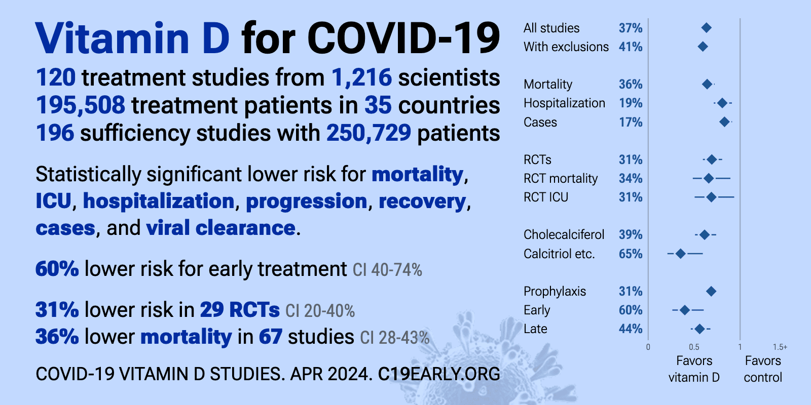
As of March 31, 2024, the Vitamin D Life COVID page had: trial results, meta-analyses and reviews, Mortality studies see related: Governments, HealthProblems, Hospitals, Dark Skins, All 26 COVID risk factors are associated with low Vit D, Fight COVID-19 with 50K Vit D weekly Vaccines Take lots of Vitamin D at first signs of COVID 166 COVID Clinical Trials using Vitamin D (Aug 2023) Prevent a COVID death: 9 dollars of Vitamin D or 900,000 dollars of vaccine - Aug 2023
5 most-recently changed Virus entries
- The above image is automatically updated
Vitamin D Life - Vitamin D Receptor activation can be increased in many ways
Resveratrol, Omega-3, Magnesium, Zinc, Quercetin, non-daily Vit D, Curcumin, Berberine, intense exercise, Butyrate Sulforaphane Ginger, Essential oils, etc Note: The founder of Vitamin D Life uses 10 of the 16 known VDR activators
Diseases associated with a poor Vitamin D Receptor
Increased risk associated with a poor Vitamin D Receptor
Note: Some diseases reduce VDR activation
those with a * are known to decrease activation
There have actually been2701 visitors to this page since it was originally made COVID kids were more likely to have a poor VDR (4.3 X), than low Vitamin D (2.6 X) – Sept 2022Printer Friendly Follow this page for updates2504 visitors, last modified 05 Jan, 2024, This page is in the following categories (# of items in each category)Attached files
ID Name Uploaded Size Downloads 18410 COVID D and VDR_CompressPdf.pdf admin 11 Sep, 2022 291.20 Kb 228 18409 Kids COVID.jpg admin 11 Sep, 2022 28.29 Kb 296